If you notice a new or changing spot on your skin, you may wonder whether it could be skin cancer. A skin cancer diagnosis involves more than simply looking at a mole or growth. Doctors use a combination of skin examinations, medical history, and laboratory tests to determine whether a suspicious spot is cancerous.
A dermatologist may examine the affected area using specialised equipment and recommend a skin biopsy if further investigation is needed. If skin cancer is confirmed, additional tests may help determine its type, depth, and whether it has spread.
Understanding how skin cancer is diagnosed can help you know what to expect during an appointment and why each step matters.
How is skin cancer diagnosed?
Skin cancer diagnosis typically involves several steps, depending on the appearance of the suspicious spot and the type of cancer being investigated.
The process may include:
- Skin examination: A doctor examines the suspicious spot and may check the rest of your skin for other unusual changes.
- Medical history: Your doctor asks about changes to the spot, previous skin conditions, sun exposure, and personal or family history of skin cancer.
- Skin biopsy: If a spot looks suspicious, a doctor removes some or all of the affected tissue for laboratory examination.
- Further tests and staging: If cancer is confirmed, additional investigations may be recommended to determine its extent and whether it has spread.
A biopsy is generally needed to confirm a skin cancer diagnosis. Visual examination alone cannot reliably establish whether a suspicious lesion is cancerous.
Skin examination
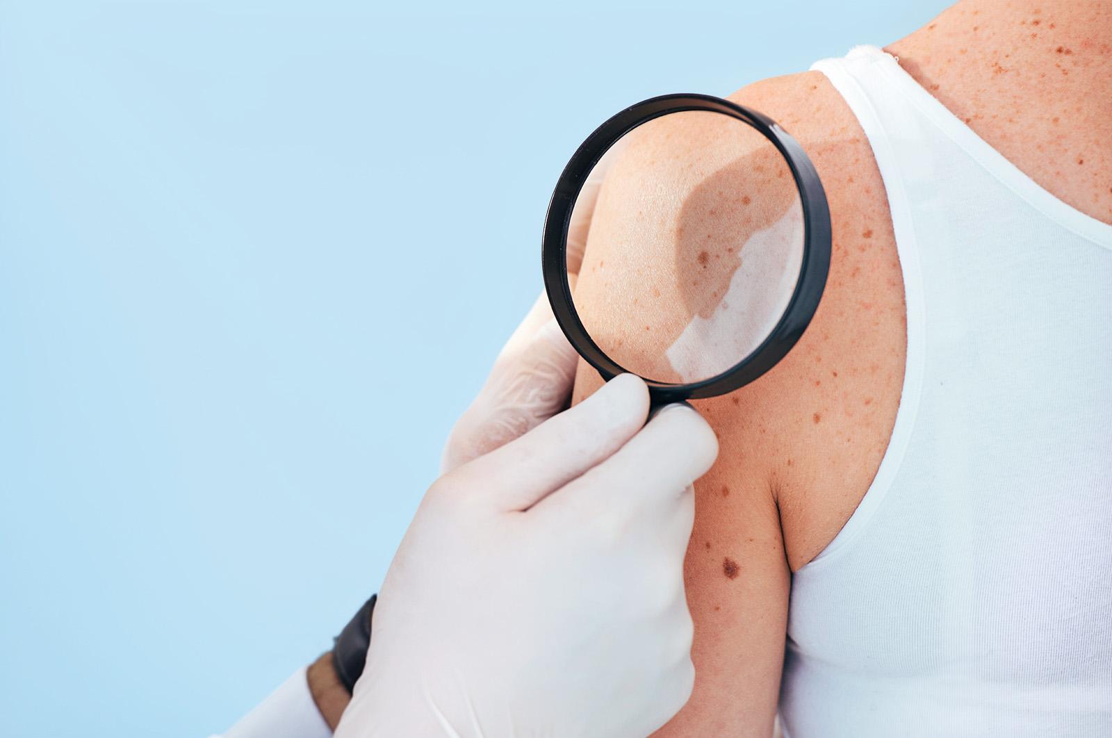
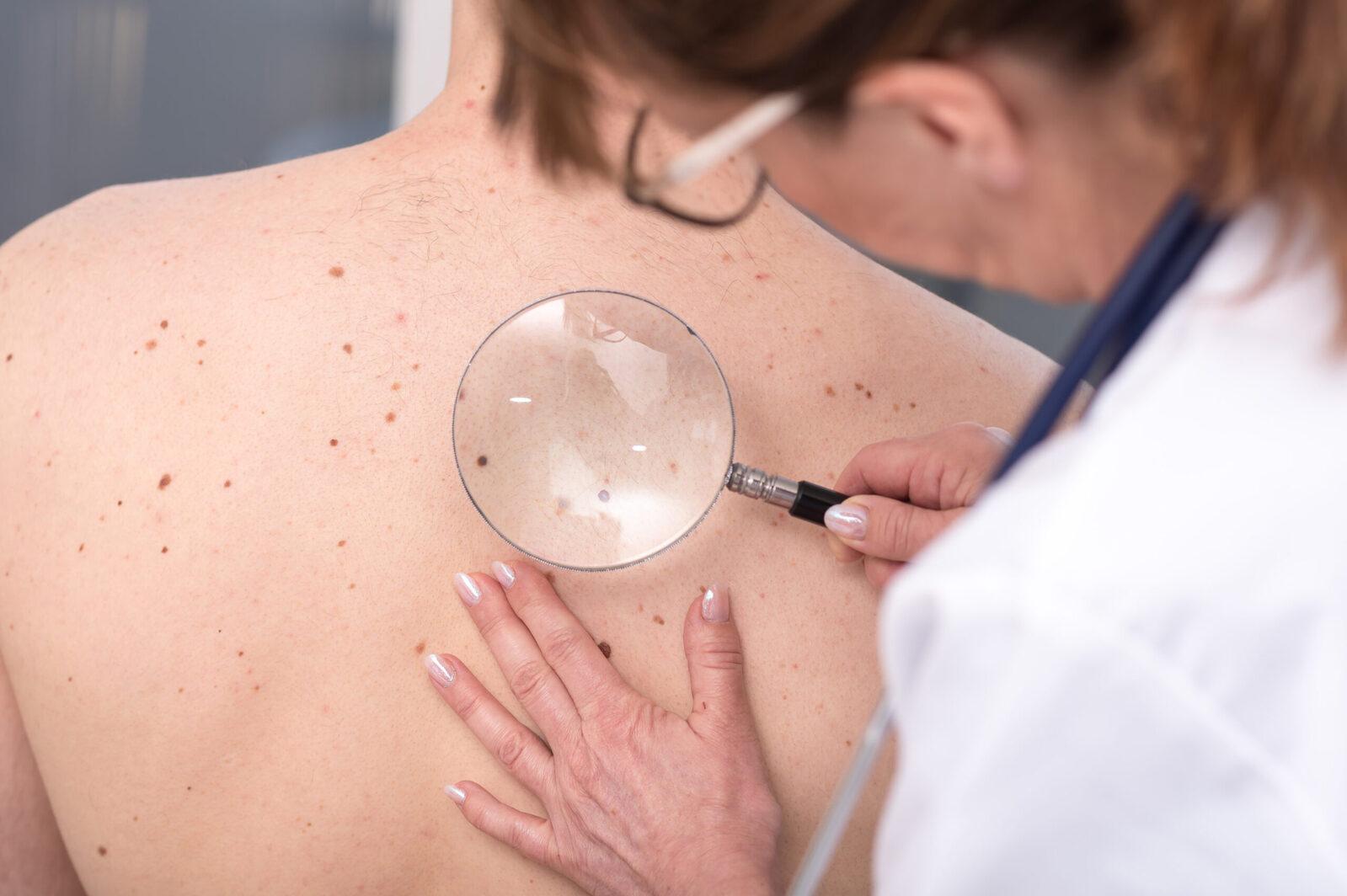
The first step in skin cancer diagnosis is usually a physical examination by a healthcare professional, such as a GP or dermatologist.
Your doctor will examine the suspicious spot, looking at its size, shape, colour, texture, and other characteristics. They may also ask when you first noticed it and whether it has changed over time.
Depending on your concerns and medical history, the doctor may recommend a more comprehensive, full-body skin examination. This allows them to check other areas for unusual moles, spots, or growths.
What is dermoscopy?
A dermatologist may use a handheld device called a dermatoscope to examine a suspicious skin lesion more closely.
Dermoscopy provides magnified views of structures and patterns beneath the skin’s surface that may not be visible to the naked eye. It can help a dermatologist assess whether a spot needs further investigation.
However, dermoscopy does not replace a biopsy when tissue confirmation is required.
Skin biopsy
If a doctor suspects that a skin lesion could be cancerous, they may recommend a skin biopsy.
A skin biopsy involves removing a small sample of skin tissue, or sometimes the entire suspicious lesion, so it can be examined under a microscope by a specialist in a laboratory.
This is the key step used to confirm whether skin cancer is present and identify the type of cancer.
What happens during a skin biopsy?
A skin biopsy is often performed during an outpatient appointment. The procedure generally involves:
- Cleaning the area: The doctor cleans the skin around the suspicious lesion.
- Applying local anaesthesia: The area is numbed to minimise discomfort during the procedure.
- Removing the tissue: The doctor removes a sample or the entire lesion, depending on the suspected condition and biopsy approach.
- Sending the sample for analysis: The tissue is sent to a laboratory for examination under a microscope.
The procedure and recovery depend on the type of biopsy and the location of the lesion. Your doctor will explain how to care for the area afterwards.
Types of skin biopsy
The appropriate biopsy method depends on the lesion’s appearance, location, and suspected skin cancer type.
Common methods include:
- Shave biopsy: Removes the upper layers of a skin lesion using a small blade.
- Punch biopsy: Uses a circular instrument to remove a small, deeper section of skin.
- Excisional biopsy: This is the preferred technique when melanoma is suspected. If melanoma is confirmed, a second procedure called a wide local excision is usually carried out to remove a larger area of skin around the original site.
- Incisional biopsy: Removes only part of a larger lesion for laboratory examination.
Laboratory examination and pathology results
After the biopsy, the tissue sample is examined under a microscope by a pathologist or dermatopathologist. This examination helps determine whether the lesion is cancerous and, if so, which type of skin cancer is present.
The pathology report may include information such as:
- Whether cancer cells are present.
- The type of skin cancer identified.
- The thickness or depth of the tumor, when relevant.
- Whether the edges of the removed tissue (margins) contain cancer cells.
- Other features that may help determine the next steps in treatment.
For melanoma, tumor thickness (also called Breslow thickness) is particularly important because it helps guide staging and treatment decisions. Your doctor will explain the findings and whether further procedures or investigations are necessary.
Further tests to determine whether skin cancer has spread
Not everyone diagnosed with skin cancer requires further testing.
If the biopsy confirms skin cancer, your doctor will consider the cancer type, size, depth, location, and other risk factors to decide whether further investigations are appropriate.
These tests may include:
Lymph node examination
Your doctor may examine nearby lymph nodes to check for swelling or other changes that could suggest the cancer has spread. This may be particularly relevant for certain melanomas and higher-risk squamous cell carcinomas.
Sentinel lymph node biopsy
For selected patients with melanoma, a sentinel lymph node biopsy may be recommended.
The sentinel lymph node is the first lymph node, or group of nodes, that cancer cells are most likely to reach when spreading from the original tumour.
During this procedure, the medical team identifies and removes the sentinel lymph node so it can be examined for cancer cells. The results can help determine the stage of melanoma and guide treatment decisions.
This procedure is not for every person diagnosed with melanoma.
Imaging tests
Depending on the cancer type and suspected extent, doctors may recommend imaging tests such as:
- Ultrasound: May be used to examine nearby lymph nodes.
- CT scan: Creates detailed images of internal structures to investigate possible spread.
- MRI scan: Provides detailed images of certain areas of the body.
- PET scan: May be used in selected cases to assess areas of possible cancer activity.
These tests are generally considered when there is a clinical reason to investigate possible spread. They are not routinely required for every skin cancer diagnosis, particularly for many early-stage skin cancers.
Understanding skin cancer stages
If skin cancer is confirmed, your doctor may determine its stage to understand how extensive the disease is. Staging describes the extent of cancer in the body and helps doctors plan appropriate treatment.
For melanoma, staging generally considers factors such as tumour thickness, ulceration, lymph node involvement, and whether the cancer has spread to distant parts of the body.
Melanoma is commonly classified into four main stages:
- Stage 0: Abnormal cancer cells are confined to the outermost layer of the skin. This is also called melanoma in situ.
- Stage I: The melanoma is relatively thin and has not spread to nearby lymph nodes or distant organs.
- Stage II: The melanoma is thicker and/or has certain higher-risk features, but there is no evidence of spread to nearby lymph nodes or distant organs.
- Stage III: The melanoma has spread to nearby lymph nodes or nearby skin or tissue.
- Stage IV: The melanoma has spread to distant parts of the body.
The staging system for basal cell carcinoma and squamous cell carcinoma differs from melanoma. Not every type of skin cancer is classified in the same way.
Does every suspicious mole need a biopsy?
No. Not every unusual mole, spot, or skin growth requires a biopsy.
Dermatologists use their clinical experience, physical examination, and sometimes dermoscopy to assess whether a lesion needs further investigation.
However, you should arrange a medical assessment if you notice a new or changing spot, a mole that looks different from your others, or a growth that repeatedly bleeds, crusts, or does not heal.
Do not rely on appearance alone to rule out skin cancer. If a doctor recommends a biopsy, they can explain why it’s needed and what to expect.
Can an app diagnose skin cancer?

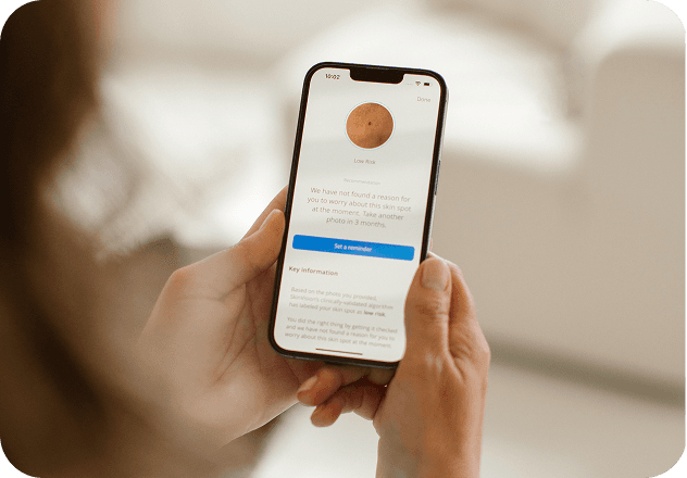
Skin-checking apps can offer an additional way to monitor suspicious spots and encourage people to seek medical advice. However, they should not be considered a replacement for a professional skin examination or biopsy.
SkinVision is a CE-marked medical device (EU MDR Class IIa) that provides a risk indication (low or high) for a skin spot, quality-checked by dermatologists, along with a recommendation to monitor the spot or see a doctor.
An app assessment does not confirm or rule out skin cancer as it also has some limitations; image quality and lighting can affect results, and the app isn’t intended for every body area or type of lesion. If you notice a suspicious or changing lesion, consult a healthcare professional even if an app assessment does not flag it as concerning.
Final thoughts
Getting a skin cancer diagnosis involves several steps, from an initial skin examination to a biopsy and, when necessary, additional tests to determine the extent of the disease. While a suspicious spot does not automatically mean cancer, changes in your skin should not be ignored.
If you notice a new, unusual, or changing skin spot, arrange an assessment with a healthcare professional. A skin cancer app like SkinVision can help you monitor spots between appointments and decide when to seek advice, but it does not replace a medical examination.
Early evaluation can help identify skin cancer sooner and ensure you receive appropriate care. Get SkinVision.
FAQs about Skin Cancer Diagnosis
How do doctors know if a spot is skin cancer?
Doctors initially examine the spot and assess its appearance, size, shape, colour, and changes over time. If they suspect skin cancer, they may perform a skin biopsy. A laboratory examination of the tissue is generally needed to confirm the diagnosis.
Can skin cancer be diagnosed without a biopsy?
A doctor may identify a lesion as suspicious through a physical examination or dermoscopy. However, a biopsy is generally needed to confirm whether it is skin cancer and identify its type.
Is a skin biopsy painful?
A local anaesthetic is usually applied before a skin biopsy to numb the area. You may feel a brief injection or some pressure during the procedure, but significant pain is not usually expected. Some tenderness may occur as the area heals.
How long does it take to get skin cancer biopsy results?
The waiting time is typically around 1-2 weeks but can vary depending on the laboratory, type of biopsy, and whether additional analysis is required. Your healthcare provider should explain when you can expect the results and how you will receive them.
What happens if a skin biopsy confirms cancer?
Your doctor will review the pathology report and discuss the next steps. Depending on the cancer type and findings, treatment may involve removing the remaining cancerous tissue or other procedures. Additional tests may be recommended if there is a concern that the cancer has spread.
Does a skin cancer diagnosis mean the cancer has spread?
No. A skin cancer diagnosis does not automatically mean the cancer has spread to other parts of the body. Many skin cancers are diagnosed while still localised to the skin. Further investigations are only recommended when clinically appropriate.